GOGGLES HELP SURGEONS SEE CANCER CELLS GLOWING LIVE DURING SURGERY
GOGGLES HELP SURGEONS SEE CANCER CELLS GLOWING LIVE DURING SURGERY.
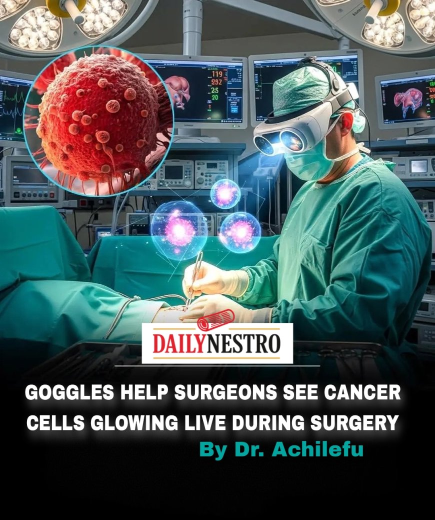
New goggles that allow surgeons to see cancer cells during an operation are a form of fluorescence-guided surgery (FGS). This technology works by combining a special dye with a sophisticated imaging system, often integrated into a wearable device like goggles. Here's a breakdown of how it works:
1. The Fluorescent Dye (Fluorophore)
The process begins with the patient being injected with a special dye, known as a fluorophore. This isn't just any dye; it's designed to specifically target and attach to cancer cells. These dyes can be engineered in several ways:
- They may contain proteins or molecules that are attracted to the unique properties of tumor cells.
- They might respond to the specific microenvironment of a tumor.
- Some newer research is even exploring bacteria engineered to emit a fluorescent signal when they encounter tumor tissue.
Once injected, the dye circulates through the patient's body and accumulates in the cancerous areas. The key is that the dye itself does not glow on its own.
2. The Goggles and Imaging System
The "goggles" are a head-mounted device equipped with a specialized camera and a light source, often a near-infrared (NIR) laser. Here's what happens during the operation:
- Activation: The surgeon activates the goggle system. A laser light shines across the patient's tissue.
- Emission: This laser light excites the fluorescent dye that has attached to the cancer cells. This causes the dye to emit a unique, invisible light signal, typically in the near-infrared spectrum.
- Detection: The camera in the goggles is specifically sensitive to this near-infrared light. It detects the signal from the glowing cancer cells.
- Augmented Reality Overlay: The system's processor takes the invisible light signal and converts it into a visible image. This image is then superimposed as a real-time, computer-generated overlay on the surgeon's view of the patient's anatomy. The cancer cells "light up," often in a distinct color like red or purple, while healthy tissue remains in its natural color.
Why This Technology Is a Game-Changer
- Enhanced Precision: The human eye cannot distinguish between microscopic cancer cells and healthy tissue. This technology allows surgeons to see the exact boundaries of a tumor, ensuring they can remove all cancerous tissue while sparing as much healthy tissue as possible. This is particularly important for achieving "clean margins" (areas free of cancer cells), which reduces the risk of recurrence.
- Real-Time Visualization: Unlike pre-operative imaging like CT or MRI scans, this technology provides real-time guidance during the surgery itself.
- Deeper Tissue Penetration: By using near-infrared light, which can penetrate deeper into tissue than visible light, the goggles can detect tumors that are beneath the surface, a feat that would be impossible with the naked eye.
- Reduced Cost and Size: Researchers are developing systems that are more portable, less expensive, and more user-friendly than older, bulky imaging systems, making this technology more accessible to a wider range of hospitals.
Read more on about the development.